I have probably mentioned this before, but I owe a significant debt of gratitude to all of those folks over the years who took the time to teach me. Whether it was my first and second grade teacher at Southeast Elementary or the clinic supervisors at Lutheran Medical Center, each and every teacher throughout my education has made a meaningful impact on my life. If you have an opportunity to teach, whether it is in dentistry or not, it would behoove you to take the opportunity. You will have the opportunity to shape someone else’s life in a positive way, as so many have done for you. With that said, I was asked by an instructor of mine several years ago:
“Do you like surprises?”
Seems like a strange question, but it was in response to a query of whether or not we should take an additional radiograph on a patient many years ago. It also serves as a foundational value in my everyday practice. It is the reason I still strive to adequately diagnose each case preoperatively, and also why I feel so strongly about the use of multiple preoperative radiographs. But no one gets it right all the time. In this case, a young patient was referred with a necrotic front tooth. In the interest of brevity, here is the preoperative radiograph. Please note that this is the only preoperative radiograph that was taken.
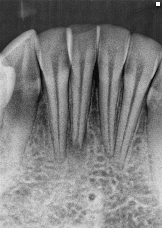
Based on Clinical and Radiographic evaluation, the diagnosis is:
Necrotic pulp, Asymptomatic apical periodontitis, tooth #25.
As it so happened, I was able to inspect the internal anatomy of the tooth under the microscope, so the final radiograph wasn’t a surprise to me. However, that’s a little bit like being saved by an airbag when you weren’t wearing a seat belt. The point is this: Treatment worked out but it was only because I was able to avoid a problem that I didn’t even know was there before I got started.
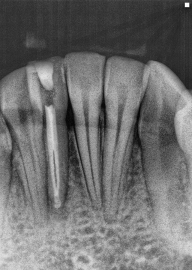
In a vital tooth, the missed canal may have been more noticeable. A failure to debride the missed canal space would likely show continuous bleeding, suggesting another portal of exit. But this tooth was nonvital. Perhaps my knowledge of the literature should have suggested this as it is a common anatomical variant. But I walked in the room, saw a young anterior tooth, and got ahead of myself. Would you have done the same? Food for thought.
A tip: For those patients who are very difficult to get radiographs on, don’t be afraid to do your diagnostics off of your initial radiograph. Additional radiographs can then be taken once the patient is comfortably numb.
A second tip: Focus on a cingulum sparing access in anterior teeth. This will move you towards the incisal edge, idealizing your angle of approach to the root canal system and improving your visualization of the internal anatomy.
Also, todays digital radiography uses less radiation than ever before. I subscribe to the concept of ALARA (As Low As Reasonable Achievable), but we need adequate radiographs to do our job. My practice is no different from yours in that some patients dislike radiographs for a host of reasons. However, cutting corners fulfills no ones objectives in the end. Does anyone argue with the concept of Call Before You Dig when a construction project is starting? I think not.
And to answer the question:
“No, I don’t like surprises, not even on my birthday. And especially not in the operatory.”
See you in 2016!